Understanding emphysema
Emphysema is part of a group of diseases called Chronic Obstructive Pulmonary Disease (COPD) that also includes chronic bronchitis. People with COPD have damage to their lungs, making it difficult for air to get in and out interfering with the lungs’ ability to deliver oxygen to vital organs.
Emphysema is a chronic lung condition caused by the damage to the alveoli, the tiny air sacks in the lungs where the exchange of oxygen and carbon dioxide occurs. This leads to breathlessness, the main symptom of emphysema. In addition to compromised gas exchange, the damage to the alveoli leads to the loss of lungs’ elasticity and results in air getting trapped inside the lungs. This makes the lungs abnormally large, further exacerbating the feeling of breathlessness.
This is why some of the treatments for emphysema are aimed at lung volume reduction, for instance endobronchial valve treatment

As people get older, the lungs slowly lose function. In people with emphysema, the muscles that help them breathe tire more quickly leading to the feeling of breathlessness with even the slightest of activities. As symptoms progress, people with emphysema may feel breathless even when at rest.
Symptoms range from mild to very severe and may also include the following:
- Shortness of breath
- Chronic cough
- Increased mucus production
- Wheezing and chest tightness
- Fatigue
- Loss of appetite and weight
- Anxiety and depression
The most significant cause of emphysema is cigarette smoking. It is also the most preventable cause. Other common risk factors include air pollution, passive smoking, work related exposure to dust and other occupational pollutants and genetic disorders, such as alpha-1-antitrypsin deficiency. Older age is a significant risk factor for emphysema and is likely to become even more relevant in the future as our population ages.
Frequent exposure to cigarette smoke leads to destruction of lung tissue and also causes inflammation of airways. Eventually this results in air flow obstruction. Cigarette smoke destroys the protective lining of airways, which means that mucous secretions cannot be cleared effectively. At the same time, mucus production increases, which can lead to respiratory infections. Air pollution has a similar role in development of the disease as it causes inflammation of airways and destruction of lung tissue.
Alpha-1-antitrypsin deficiency is a rare disorder and the only known genetic factor that increases the risk of developing emphysema. Over time, people with alpha-1-antitrypsin deficiency develop emphysema because they are unable to fight a destructive enzyme in the lungs called trypsin.
Trypsin is a digestive enzyme, most often found in the digestive tract, where it is used to help the body digest food. It is also released by immune cells in their attempt to destroy bacteria and other material. The destruction of tissue by trypsin produces similar effects to those seen with cigarette smoking.
How is emphysema diagnosed?
 You may have difficulty breathing or have a persistent cough, but these symptoms alone do not mean that you have emphysema. On the other hand, many people who have emphysema may not be diagnosed until the disease is quite advanced and is therefore more difficult to treat. Your doctor may perform a number of tests to diagnose emphysema and determine the severity of the disease.
Physical examination
In the first instance, your doctor will ask you some specific questions about your general state of health and your condition, as well as conduct a general physical examination. They will mainly focus on your heart and lungs by measuring your blood oxygen levels, assessing the rate and pattern of your breathing and by listening to the breathing sounds of your chest.
Lung function tests
There are a variety of lung function tests that are useful in diagnosing emphysema. These tests are designed to measure how well the lungs are moving air in and out and also to estimate how well the lungs are delivering oxygen to the blood.
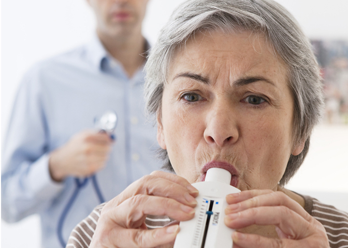
Spirometry is the most common lung function test. It objectively measures the degree of airflow limitation caused by narrowing of the airways as a result of COPD. It is a non-invasive and readily available test. Spirometry is used to detect emphysema as well as track the progression of the disease and the effectiveness of treatment.
The test is performed in the clinic and involves inhaling fully and then breathing all the way out into a device called a spirometer. This provides information about the flow and volume of air entering the lungs.
Spirometry measures the total volume of air exhaled from the point of maximal inspiration (forced vital capacity, FVC) and the volume of air exhaled during the first second of exhalation (forced expiratory volume in one second, FEV1). The ratio of these two measurements (FEV1/FVC) can also be calculated and is necessary to diagnose COPD.
This is another lung function test that is particularly useful in providing additional information about the disease. It can measure the amount of air in your lungs after you take a deep breath. It also measures the amount of air left in your lungs after full exhalation. In patients with emphysema, this amount of residual air can be significantly larger due to the air getting trapped in the lungs.
This test is particularly important to perform if lung volume reduction is being considered as a possible treatment.
Another measurement of lung function, which is easy to perform, is the carbon monoxide transfer test. It helps to estimate how well the lungs are performing their most basic and most important function: getting oxygen into the blood stream.
Arterial blood gas analysis measures the levels of oxygen and carbon dioxide in the blood from an artery. This is generally used for patients with severe disease and those being considered for oxygen therapy.
Exercise testing allows lung and heart function to be measured under the stress of exercise. Cardiopulmonary exercise tests may be useful to differentiate between breathlessness resulting from cardiac or respiratory disease, and may help to identify other causes of exercise limitation. Walking tests such as the 6-minute walking test evaluates the distance you can walk in 6 minutes and provides an indication about your exercise capacity.
Imaging tests
Chest x-ray is often done but it is not sensitive for the diagnosis of emphysema. It is mainly done to exclude other diseases such as lung cancer.
Providing detailed images of the lung structures, high resolution computed tomography (HRCT) scanning can help diagnose the presence of emphysema and determine its type and extent.
If emphysema is diagnosed, HRCT scans can be further analysed using specialised software to give physicians even more useful information. For instance, it can accurately predict the likelihood that patients will respond to certain types of treatments, such as lung volume reduction with endobronchial valves.
The ventilation and perfusion scan is used to measure the circulation of air and blood in the lungs. This helps your doctor to evaluate how much blood flows to different parts of your lungs. This is important to know because if blood flows to the areas damaged by emphysema, the exchange of oxygen and carbon dioxide may be impaired. These scans also help assess whether patients would benefit from surgical or endoscopic lung volume reduction.
